Table of Contents
Part I: The Case That Broke Me: When “Routine” Back Pain Becomes a Life Sentence
Introduction: The Ghost in the Exam Room
In the early years of my career as a medical researcher, I carried the confident, slightly naive belief that our diagnostic systems were robust. We had protocols, algorithms, and a wealth of collective knowledge. Then I met “Mr. Harrison,” a name I’ve created for a man whose real story has haunted my work ever since. He was a retired accountant, a man of numbers and quiet routines, who came to the emergency department with severe, unyielding lower back pain. He had no history of trauma. His initial lab work was largely unremarkable. He was diagnosed with mechanical back pain, given analgesics, and sent home with instructions for rest. It was a common presentation with a common diagnosis. It was routine.
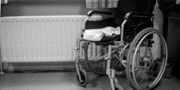
The catastrophe came weeks later. Mr. Harrison returned, not walking, but wheeled in on a gurney. The pain had become unbearable, and now it was accompanied by a terrifying new symptom: he couldn’t feel his legs. The diagnosis, when it finally came, was a massive, untreated spinal epidural abscess. The collection of pus had been silently growing in his epidural space, compressing his spinal cord until the damage was irreversible. Despite emergency surgery, it was too late. The delay had cost him his mobility, leaving him with permanent, incomplete tetraplegia.1 He awoke from the ordeal unable to stand or walk, his life irrevocently altered by a “routine” complaint that was anything but.1
That failure was more than a missed diagnosis; it was a catastrophic failure of the diagnostic system itself. It exposed a fatal flaw in how we approach common symptoms. The case became the central, motivating struggle of my professional life. It left me with a haunting question that has driven every research project since: How do we see the monster hiding in plain sight? How do we catch the culprit responsible for these tragedies before it destroys a life? The stakes are not just academic; they are measured in futures lost and families devastated.1
Part II: The Forensic Epiphany: A New Way of Seeing
From Clinician to Investigator
The weight of that failure led to a period of deep professional disillusionment. The standard models had failed. I began searching for new methodologies outside the confines of traditional medicine, looking for a more rigorous, more urgent way to approach diagnostic uncertainty. The epiphany arrived from a field that seemed, at first, entirely unrelated: forensic architecture and engineering.5
Forensic investigators operate on a fundamental principle articulated by the pioneering criminologist Edmond Locard and echoed in modern forensic science: every contact leaves a trace.6 When they approach a crime scene, they don’t assume anything. They meticulously gather evidence, document every detail, and reconstruct the event to understand what happened. I realized this was the exact mindset needed in the clinic.
This led to the development of a new framework. In this model, the patient’s body is the “crime scene.” Their symptoms and history are the “witness testimony.” The lab results and imaging studies are the “physical evidence.” The underlying disease is the “culprit.” The clinician’s role transforms from a passive treater of symptoms to an active, impartial forensic investigator. The goal is not merely to manage the patient’s complaint but to meticulously reconstruct the timeline of the “crime,” identify the perpetrator, and intervene before irreversible damage occurs.7
This forensic mindset is a powerful antidote to the clinical inertia that so often leads to diagnostic delays. Back pain is extraordinarily common, affecting up to 85% of adults at some point in their lives.8 In contrast, spinal infections are rare, accounting for only 3% to 5% of all osteomyelitis cases, which are themselves uncommon.9 This statistical reality creates a powerful cognitive bias, predisposing clinicians to default to a benign diagnosis. The forensic framework shatters this bias. It forces a fundamental shift in the guiding question from, “What is the most likely diagnosis?” to a more urgent and responsible one: “What is the most dangerous diagnosis I must prove is
not present?” It demands rigor, impartiality, and a systematic approach, just like a real forensic investigation.7
Part III: The Forensic Framework: A Step-by-Step Investigation of the Spine
Step 1: Recognizing a Potential Crime Scene (The Red Flag System)
The first duty of an investigator arriving at a scene is to assess the situation. In clinical terms, this means determining whether the case is a “civil dispute” (e.g., simple mechanical back pain) or a potential “felony” (a serious underlying condition requiring urgent action). The Red Flag System is the protocol for making this critical determination. The presence of any of these flags should immediately trigger a full-scale investigation.
The key red flags for a potentially serious spinal pathology include:
- Constitutional Symptoms: The presence of systemic signs like fever, chills, or unexplained weight loss is a major warning. While fever is present in only 35% to 60% of vertebral osteomyelitis cases, its appearance with back pain is highly suspicious.9
- Pain Characteristics: The nature of the pain is a crucial clue. Unlike mechanical pain, which often improves with rest, pain from an infection or tumor is frequently severe, unrelenting, and may even worsen at night or when lying down.12
- Neurological Deficits: Any new or progressing neurological symptom is an emergency signal. This includes weakness, numbness, or tingling in the arms or legs, or signs of cauda equina syndrome, such as loss of bladder or bowel control and “saddle anesthesia” (numbness in the groin and buttocks).14
- Patient History (Risk Factors): The patient’s background can significantly elevate suspicion. Key risk factors include a recent bacterial infection (e.g., urinary tract, skin, or dental infection), a recent spinal procedure like surgery or an epidural injection, a history of intravenous drug use, or any condition that compromises the immune system, such as diabetes, HIV, cancer, or long-term steroid use.9
The overwhelming prevalence of benign back pain creates a “diagnostic inertia” that is difficult to overcome. The Red Flag System acts as a circuit breaker for this cognitive bias. It provides a non-negotiable checklist that, if positive, compels the clinician to abandon the default diagnosis and escalate to an investigative footing.
Table 1: The Forensic Investigator’s Red Flag Checklist
| Category | Red Flag | Potential Serious Cause(s) |
| Patient History | Age > 50 years with new onset pain | Cancer, Fracture, Infection 12 |
| History of Cancer | Metastasis 12 | |
| Unexplained Weight Loss | Cancer, Infection 14 | |
| Immunosuppression (Diabetes, Steroids, HIV) | Infection 9 | |
| Intravenous Drug Use | Infection (especially Epidural Abscess) 9 | |
| Recent Bacterial Infection (UTI, Skin) | Infection (Osteomyelitis, Discitis) 3 | |
| Recent Spinal Procedure or Surgery | Infection 3 | |
| Pain Characteristics | Severe, Unrelenting Pain | Infection, Cancer 14 |
| Pain Worse at Night or at Rest | Infection, Cancer 12 | |
| No Improvement with Rest | Inflammatory Cause (Infection) 12 | |
| Neurological Signs | Progressive Motor or Sensory Deficit (Weakness, Numbness) | Spinal Cord/Nerve Root Compression 12 |
| Saddle Anesthesia | Cauda equina syndrome 12 | |
| Bladder or Bowel Incontinence/Retention | Cauda equina syndrome 13 | |
| Constitutional Symptoms | Fever, Chills | Infection 14 |
Step 2: Securing the Scene & Gathering Initial Evidence (The Diagnostic Toolkit)
Once red flags indicate a potential crime scene, the investigation must begin immediately. Delay is the primary accomplice to tragedy in these cases.4 The first steps involve gathering initial evidence from the scene (the patient).
- Gathering “Testimony” (Patient History): In the forensic framework, the patient’s story is not just a formality; it is crucial witness testimony that guides the entire investigation. Forensic architecture principles value witness accounts as a primary tool for locating and interpreting physical evidence.5 Patient forums are filled with stories of individuals whose severe pain was dismissed because initial tests were normal, highlighting a desperate need to be believed.21 An investigator must listen intently, probing for the high-risk factors identified in the research: diabetes, immunosuppression, IV drug use, recent surgeries, or any prior infections, no matter how seemingly minor.9 A strong “testimony” (severe, unyielding pain plus risk factors) demands a search for corroborating physical evidence, even if initial labs are ambiguous.
- Collecting “Trace Evidence” (Lab Work): The next step is to collect objective physical evidence through blood work.
- Inflammatory Markers: The two most important initial blood tests are the Erythrocyte Sedimentation Rate (ESR) and C-reactive protein (CRP). These markers of inflammation are highly sensitive for spinal infection, with elevations found in over 95% of cases.10 An elevated ESR or CRP in a patient with red flags is a major piece of evidence demanding further investigation.
- Complete Blood Count (CBC): While a CBC is standard, it is critical to understand its limitations. The white blood cell count can be normal in a significant number of patients with confirmed spinal infections, making it an unreliable marker for ruling out disease.10 Relying on a normal WBC for reassurance is a common and dangerous investigative error.
- Blood Cultures: Before any antibiotics are administered, at least two sets of blood cultures must be drawn.9 These cultures can identify the causative organism in a large percentage of cases, providing the “identity of the perpetrator” and allowing for precisely targeted antibiotic therapy.9
Step 3 & 4: Analyzing the Evidence & Profiling the Suspects (Differential Diagnosis)
With initial evidence in hand, the investigator develops a list of “prime suspects.” In the context of spinal infections, there are three main culprits, each with a distinct profile or “rap sheet.”
- Suspect Profile 1: Vertebral Osteomyelitis (The Bone Invader)
- Modus Operandi: This is an infection of the vertebral bone itself, most often caused by bacteria like Staphylococcus aureus that travel through the bloodstream (hematogenous spread) from an infection elsewhere in the body.9
- Victimology (Location): It has a clear preference for the lumbar spine (58% of cases), followed by the thoracic (30%) and cervical (11%) regions.9
- Clinical Clues: Its presentation is notoriously nonspecific. Back pain is the most common symptom, but fever is often absent, and tenderness to the touch over the spine is only present in a minority of patients.9 This stealthy nature makes it easy to misdiagnose as a simple strain.
- Suspect Profile 2: Discitis (The Cushion Corruptor)
- Modus Operandi: This is an infection of the soft, cushion-like intervertebral disc space, often occurring alongside osteomyelitis (a combined condition called spondylodiskitis).26 The disc’s poor blood supply is a key vulnerability; it makes it difficult for the body’s immune cells and antibiotics to reach the infection, allowing it to fester.10
- Victimology (Demographics): It typically affects two distinct populations: young children and older adults, with an increased incidence around age 50.10
- Clinical Clues: Severe, debilitating, and localized back pain is the hallmark symptom.10 It can also be accompanied by abdominal pain, stiffness, and noticeable changes in posture.26
- Suspect Profile 3: Spinal Epidural Abscess (SEA) (The Silent Strangler)
- Modus Operandi: This is a collection of pus in the epidural space, the area surrounding the spinal cord. It is the most urgent and dangerous of the three, as the abscess can physically compress the spinal cord and its nerve roots, leading to paralysis.15 It is a true neurosurgical emergency.
- Victimology (Risk Factors): There is a strong association with IV drug use, diabetes, recent spinal procedures, and other sources of bacteremia.11
- Clinical Clues: The “classic triad” of back pain, fever, and neurologic deficit is a diagnostic trap; it is present in only 8-15% of cases.11 The disease is known to progress through four stages, often with terrifying speed. A patient can move from Stage 3 (sensory and motor deficits) to Stage 4 (complete paralysis) in less than 24 hours.16 This rapid progression underscores the absolute urgency of the investigation.
Table 2: Comparing the Culprits: A Forensic Profile of Spinal Infections
| Feature | Vertebral Osteomyelitis | Discitis | Spinal Epidural Abscess (SEA) |
| Primary Location | Vertebral bone 9 | Intervertebral disc space 26 | Epidural space 15 |
| Common Location | Lumbar spine (58%) 9 | Lumbar spine 10 | Lumbar spine (48%) 16 |
| Key Demographics | Advanced age, immunosuppressed 9 | Bimodal: Young children & adults >50 10 | IV drug users, diabetics, post-procedure 16 |
| Common Pathogens | Staphylococcus aureus, E. coli 9 | Staphylococcus aureus 27 | Staphylococcus aureus 15 |
| Hallmark Symptom | Nonspecific, persistent back pain 9 | Severe, localized, debilitating back pain 26 | Severe back pain, focal tenderness 15 |
| “Red Alert” Feature | Often lacks fever or focal tenderness 9 | Poor blood supply makes treatment difficult 10 | Rapid neurological progression to paralysis 16 |
Step 5 & 6: Deploying Advanced Surveillance & Testing the Hypothesis (Definitive Imaging)
The spine is a silent, high-stakes crime scene. Its deep, complex anatomy, particularly the avascular nature of the discs, allows infection to develop without obvious external signs.10 However, the consequences of a missed infection—spinal cord compression, paralysis, and even death—are catastrophic.1 Therefore, an investigator cannot afford to wait for overwhelming evidence. Once red flags are raised and initial evidence is gathered, advanced surveillance must be deployed immediately.
- The Unblinking Eye: MRI: The single most important test in the investigation of a suspected spinal infection is Magnetic Resonance Imaging (MRI) with and without gadolinium contrast.9 It is the undisputed gold standard.
- Why Other Imaging Fails: Plain X-rays are notoriously insensitive in the early stages of infection; findings may not appear for several weeks, creating a dangerous window of false reassurance.10 A CT scan is better than an X-ray for viewing bone but remains inferior to MRI for visualizing soft tissues, inflammation, and abscess formation.9
- What the MRI Reveals: The MRI is the high-tech surveillance tool that can see what other tests miss. It can detect bone marrow edema, inflammation in the disc space, destruction of the vertebral endplates, and, most critically, the presence of a fluid collection like an epidural abscess.9 It provides the definitive visual evidence to confirm or refute the hypothesis of a spinal infection.
Step 7: Obtaining Conclusive Proof & Identifying the Perpetrator (Biopsy and Culture)
While an MRI can confirm that a “crime” (an infection) has occurred, it cannot definitively identify the “perpetrator” (the specific pathogen). For that, the investigator needs the “smoking gun.”
- Image-Guided Biopsy: In most cases, a CT-guided needle biopsy is performed to obtain a tissue sample from the infected area.9 This sample is then sent for culture and histopathology.
- The Exception to the Rule: A biopsy may not be necessary if the patient already has positive blood cultures with a typical pathogen (like S. aureus) and MRI findings that are consistent with that infection.23
- The Importance of Identification: Obtaining a culture is crucial because it allows for the definitive identification of the causative bacteria or fungus and its specific antibiotic susceptibilities. This enables the medical team to move from broad-spectrum “dragnet” antibiotics to a targeted, “precision” therapy that is most effective against the specific invader.9
Step 8: The Verdict, Intervention, & Post-Case Follow-Up (Treatment & Management)
A forensic investigation is a team effort, requiring a range of specialists to solve the case and manage the aftermath.35 The same is true for treating spinal infections. The initial clinician acts as the “first responder” who recognizes the crime scene and sounds the alarm. Their success is measured by the speed and accuracy with which they activate the expert response team.
- Assembling the “Task Force”: Effective management requires a multidisciplinary approach. The core team includes specialists in Infectious Disease (to manage antibiotic therapy), Neurosurgery or Orthopedic Surgery (to assess spinal stability and perform surgery if needed), and Interventional Radiology (to perform biopsies).9
- The Intervention Plan (Treatment):
- Antimicrobial Therapy: This is the foundation of treatment. It involves a long course, typically at least 6 weeks, of pathogen-targeted intravenous (IV) antibiotics.9 In some cases, this may be followed by a course of oral antibiotics.33
- Surgical Intervention: Surgery is reserved for specific, urgent indications:
- Neurological Compromise: Any patient with progressive neurological deficits or evidence of spinal cord compression from an abscess requires emergency surgical decompression.9
- Spinal Instability: If the infection has destroyed bone to the point of causing spinal instability or deformity, surgery is needed to stabilize the spine, often with hardware.9
- Failure of Medical Therapy: If the infection does not respond to appropriate antibiotic therapy, surgery may be required to debride (clean out) the infected tissue.9
- Monitoring the Case: After the intervention, the case is monitored closely. Critically, the response to treatment is tracked not by repeat imaging (which can remain abnormal for a long time and be misleading), but by the patient’s clinical improvement and the normalization of their inflammatory markers (ESR and CRP).23
Table 3: Initial Empiric Antibiotic Regimens for Suspected Spinal Infections
(Note: This is a guide for initial therapy only. Treatment must be tailored to culture and susceptibility results once available.)
| Suspected Organism(s) | Preferred Regimens | Alternative Regimens | ||
| Methicillin-resistant S. aureus (MRSA) | Vancomycin, 15 to 20 mg per kg per dose IV every 8 to 12 hours | Linezolid, 600 mg IV or orally every 12 hours; Daptomycin, 6 mg per kg IV every 24 hours | ||
| Methicillin-sensitive S. aureus (MSSA) | Nafcillin or Oxacillin, 2 g IV every 4 hours; Cefazolin, 2 g IV every 8 hours | Vancomycin, 15 to 20 mg per kg per dose IV every 8 to 12 hours | ||
| Gram-negative bacilli (e.g., E. coli) | Ceftriaxone, 2 g IV every 24 hours; Ciprofloxacin, 400 mg IV every 8 to 12 hours | Piperacillin/tazobactam, 3.375 g IV every 6 hours | ||
| Pseudomonas aeruginosa | Cefepime, 2 g IV every 8 hours OR Piperacillin/tazobactam, 4.5 g IV every 6 hours | Meropenem, 1 g IV every 8 hours | ||
| 31 |
Part IV: Conclusion: A New Standard of Care
From Tragedy to Triumph: A Case Solved
I often think back to Mr. Harrison and the systemic breakdown that failed him. But I also hold onto the cases that show us a better way. I recall a more recent patient, a 55-year-old woman with poorly controlled diabetes who presented with severe thoracic back pain and a low-grade fever. The red flags were immediately recognized. Instead of being sent home, she was admitted. The forensic investigation was launched. Her ESR and CRP were highly elevated. Blood cultures were drawn, and an urgent MRI with contrast was ordered. The MRI revealed T7-T8 discitis and osteomyelitis with a small epidural fluid collection. The “task force” was activated. The infectious disease team started empiric IV antibiotics covering MRSA and gram-negatives, and the neurosurgery team was consulted. Her blood cultures grew MSSA, and her antibiotics were tailored. She never developed neurological deficits. After six weeks of targeted IV therapy, she made a full recovery. She avoided the fate of Mr. Harrison because the paradigm was different. The investigation was swift, systematic, and predicated on ruling out the worst-case scenario.
A Call for a Paradigm Shift
Spinal infections are master mimics, dangerous predators that camouflage themselves with the mundane symptom of back pain. The cost of a missed diagnosis is not measured in dollars, but in futures—in the ability to walk, to work, to live without pain or paralysis. The forensic framework presented here is not an academic exercise; it is a necessary, life-saving shift in clinical mindset.
To every healthcare professional on the front lines, the call to action is clear: Treat every case of severe, unexplained back pain with the rigor and urgency of a forensic investigator. Assume the worst until you can prove otherwise. Listen to your patient’s testimony as if it were evidence in court. Gather the physical evidence meticulously. Activate the expert task force without delay. And never forget the human life that hangs in the balance of your investigation. Our collective goal must be to build a system of vigilance and rigor that ensures there are no more “Mr. Harrisons.”
Works cited
- Spinal infection left client tetraplegic after a delay in diagnosis, accessed on August 11, 2025, https://www.stewartslaw.com/news/client-left-tetraplegic-spinal-infection/
- Discitis Negligence Case Study | Delayed Diagnosis – Medical Solicitors, accessed on August 11, 2025, https://www.medical-solicitors.com/case-studies/delayed-diagnosis-cases/missed-discitis-spinal-infection-leads-to-six-figure-compensation/
- Delayed Diagnosis of Spinal Infection – Lupetin & Unatin, LLC, accessed on August 11, 2025, https://www.pamedmal.com/delayed-diagnosis-of-spinal-infection/
- Vertebral Osteomyelitis: Long-Term Outcome for 253 Patients from 7 Cleveland-Area Hospitals | Clinical Infectious Diseases | Oxford Academic, accessed on August 11, 2025, https://academic.oup.com/cid/article/34/10/1342/327358
- Forensic Architecture – Right Livelihood, accessed on August 11, 2025, https://rightlivelihood.org/the-change-makers/find-a-laureate/forensic-architecture/
- What is Architectural Forensics? | ArchDaily, accessed on August 11, 2025, https://www.archdaily.com/983759/what-is-architectural-forensics
- 8 steps to a successful forensic investigation – EFI Global, accessed on August 11, 2025, https://www.efiglobal.com/8-steps-to-a-successful-forensic-investigation/
- Vertebral Osteomyelitis: Unexpected Cause of Back Pain | Lopez-Rivera, accessed on August 11, 2025, https://www.journalmc.org/index.php/JMC/article/view/2870/2195
- Vertebral Osteomyelitis – StatPearls – NCBI Bookshelf, accessed on August 11, 2025, https://www.ncbi.nlm.nih.gov/books/NBK532256/
- Diskitis – StatPearls – NCBI Bookshelf, accessed on August 11, 2025, https://www.ncbi.nlm.nih.gov/books/NBK541047/
- Spinal Epidural Abscess: A Review Highlighting Early Diagnosis and Management | JMA Journal, accessed on August 11, 2025, https://www.jmaj.jp/detail.php?id=10.31662%2Fjmaj.2019-0038
- Don’t Miss Red Flags for Serious Back Pain | EBSCO, accessed on August 11, 2025, https://www.ebsco.com/blogs/health-notes/dont-miss-red-flags-serious-back-pain
- is my Back Pain serious? These 10 red flags may indicate you need urgent medical care, accessed on August 11, 2025, https://www.blueridgeortho.com/post/is-it-serious-here-are-10-red-flags-for-back-pain
- What Are the Red Flags for Low Back Pain? | Microspine PLC, accessed on August 11, 2025, https://www.microspinemd.com/2024/09/01/what-are-the-red-flags-for-low-back-pain/
- Spinal epidural abscess – Symptoms, diagnosis and treatment | BMJ Best Practice US, accessed on August 11, 2025, https://bestpractice.bmj.com/topics/en-us/926
- Spinal Epidural Abscess – StatPearls – NCBI Bookshelf, accessed on August 11, 2025, https://www.ncbi.nlm.nih.gov/books/NBK441890/
- Spinal Infections – AANS, accessed on August 11, 2025, https://www.aans.org/patients/conditions-treatments/spinal-infections/
- Spinal Infections | Aurora Health Care, accessed on August 11, 2025, https://www.aurorahealthcare.org/services/aurora-spine-services/spinal-infections
- Osteomyelitis Diagnosis & Treatment – NYC | Columbia Neurosurgery in New York City, accessed on August 11, 2025, https://www.neurosurgery.columbia.edu/patient-care/conditions/osteomyelitis
- Forensic Architecture – Events | UNSW Sydney, accessed on August 11, 2025, https://www.events.unsw.edu.au/article/forensic-architecture
- Nerve pain after discitis/osteomylitis of lumbar spine – Mayo Clinic Connect, accessed on August 11, 2025, https://connect.mayoclinic.org/discussion/nerve-pain-after-discitisosteomylitis-of-lumbar-spine/
- Spinal Infections Risk Factors | Stanford Health Care, accessed on August 11, 2025, https://stanfordhealthcare.org/medical-conditions/back-neck-and-spine/spinal-infection/risk-factors.html
- 2015 Infectious Diseases Society of America (IDSA) Clinical Practice Guidelines for the Diagnosis and Treatment of Native Vertebral Osteomyelitis in Adults, accessed on August 11, 2025, https://www.idsociety.org/globalassets/idsa/practice-guidelines/2015-infectious-diseases-society-of-america-idsa-clinical-practice-guidelines-for-the-diagnosis-and-treatment-of-native-vertebral-osteomyelitis-in-adults.pdf
- Spinal Infections | Spine Care – El Camino Health, accessed on August 11, 2025, https://www.elcaminohealth.org/services/spine-care/conditions-treatments/spinal-infections
- Osteomyelitis – Symptoms and causes – Mayo Clinic, accessed on August 11, 2025, https://www.mayoclinic.org/diseases-conditions/osteomyelitis/symptoms-causes/syc-20375913
- Discitis Causes, Symptoms, and Treatments – UPMC, accessed on August 11, 2025, https://www.upmc.com/services/spine/conditions/discitis
- Spinal Disc Problems (Discitis and Back Pain Red Flags) – Patient.info, accessed on August 11, 2025, https://patient.info/doctor/neurology/spinal-disc-problems-including-red-flag-signs
- Discitis – A Severe Cause of Back Pain | MUSC Health | Charleston SC, accessed on August 11, 2025, https://muschealth.org/medical-services/geriatrics-and-aging/healthy-aging/discitis
- Discitis – Birmingham, AL – Spine and Neurosurgery, accessed on August 11, 2025, https://neurosurgicalassociatespc.com/discitis/
- Discitis Spine Disease and Treatment – Goodman Campbell, accessed on August 11, 2025, https://www.goodmancampbell.com/conditions/spine/degenerative-disease/discitis/
- Spinal Epidural Abscess – Neurologic Disorders – Merck Manual Professional Edition, accessed on August 11, 2025, https://www.merckmanuals.com/professional/neurologic-disorders/spinal-cord-disorders/spinal-epidural-abscess
- Spinal epidural abscess–experience with 46 patients and evaluation of prognostic factors – PubMed, accessed on August 11, 2025, https://pubmed.ncbi.nlm.nih.gov/12217707/
- Osteomyelitis: Diagnosis and Treatment – AAFP, accessed on August 11, 2025, https://www.aafp.org/pubs/afp/issues/2021/1000/p395.html
- Vertebral osteomyelitis: Treatment – مدیلیب, accessed on August 11, 2025, https://www.medilib.ir/uptodate/show/144958
- Architectural Forensics, accessed on August 11, 2025, https://www.decolonizing.ps/site/wp-content/uploads/2010/07/architectural-forensics_brief_final.pdf
- Osteomyelitis (Bone Infection): Causes, Symptoms & Treatment – Cleveland Clinic, accessed on August 11, 2025, https://my.clevelandclinic.org/health/diseases/osteomyelitis-bone-infection